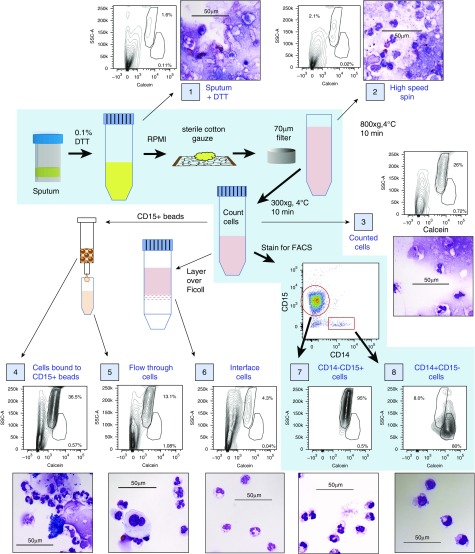
Figure 1.
Efficacy of different methods for isolating leukocytes from spontaneously expectorated cystic fibrosis (CF) sputum evaluated by flow cytometry and histologic examination. Steps in the protocol described in this article are depicted on the blue background, with thick black arrows between each step. Samples of a single specimen were evaluated at steps throughout the protocol (each sample is identified by a number in a blue box) to determine the presence of debris and the purity of the leukocyte populations by both flow cytometry and histologic examination. Thin black arrows indicate samples produced either by initial steps in the protocol or by other protocols that are commonly used to isolate leukocyte populations but are ineffective for isolating sputum leukocytes. Side scatter-area (SSC-A) versus calcein (a viability dye that fluoresces only when taken up by intact, viable cells) can be used to separate live cells and debris, and can distinguish neutrophils from macrophages more effectively than forward scatter-area (FSC-A). Larger images of histology for each sample can be found in Figure E2. Scale bars: 50 μm.

