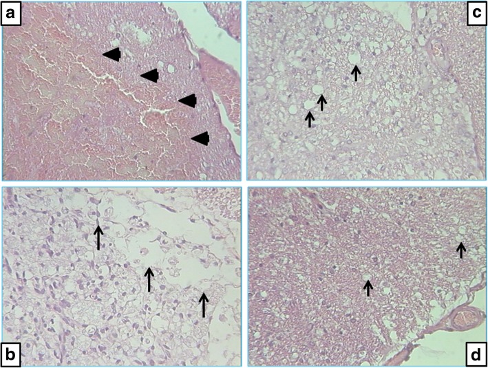
Fig. 4.
Microscopic presentation of lateral columns of rat spinal cord at the lesion site. a Ten minutes after injury, all animals have numerous fields of hemorrhage—arrowheads. b–d Histology 30 days after injury: b control animals, numerous confluent vacuoles—arrows; c BPC 157 2 μg/kg, few small vacuoles—arrows; d BPC 157200 μg/kg, only occasionally small vacuoles—arrows. Staining H&E, magnification × 300

