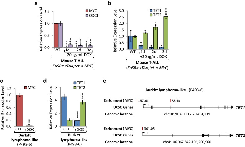
Fig. 2.
TET1 and TET2 levels are dependent on MYC expression. MYC inactivation in T-ALL cells (6780) derived from EµSRα-tTAα;tet-o-MYC mice, and human Burkitt lymphoma-like (P493-6) cells, harboring a tetracycline-regulated c-MYC allele, in a time-dependent manner for 1, 2, and 3 days using 20 ng/mL DOX. Mouse T-ALL cells: a RT-qPCR analysis of MYC and its canonical target gene Ornithine Decarboxylase 1 (ODC1), and b of TET1 and TET2. RT-qPCR data were normalized to UBC. Human Burkitt lymphoma-like cells: c RT-qPCR of MYC and c TET1 and TET2 in P493-6 cells before (CTL) and upon MYC inactivation through treatment with 20 ng/mL DOX for 2 days (+DOX). RT-qPCR data were normalized to RPL13A. d MYC ChIP-seq data for P493-6 cells obtained from Sabo et al. [13] indicating enrichment score for MYC at the TET1 and TET2 loci. Traces were generated based on reference genome hg19 using the UCSC Genome Browser. The chromosomal location is indicated in bp. MYC binding peaks are displayed as red vertical bars; numbers represent the relative fold enrichment for MYC. Exons are displayed as black vertical bars, the UTR is represented by a black line, and the transcription start site (TSS) is marked by an arrow indicating the direction of transcription

