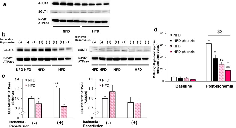
Fig. 5.
The expression of glucose transporters and the glucose uptake in myocardium during ischemia–reperfusion. Representative immunoblots of GLUT4 and SGLT1 in the plasma membrane fraction from the murine perfused hearts at baseline measured at the end of 10-min pre-ischemia perfusion (a) and before and after IRI (b) are shown. c Densitometric quantitation normalized to the level of either the GLUT4 or SGLT1 expression in NFD hearts before IRI are shown ([GLUT4] NFD or HFD without IRI: n = 8 each, NFD or HFD with IRI: n = 6 each; [SGLT1] NFD or HFD without IRI: n = 9 each, NFD or HFD with IRI: n = 7 or n = 6, respectively). *P < 0.05, **P < 0.01 versus NFD hearts before IRI; ‡P < 0.01 versus NFD hearts after IRI. In both a and b, immunoblots of Na+/K+ ATPase from the same membrane are shown as a loading control for the membrane fraction. d Glucose uptake in NFD (open black square), NFD phlorizin-perfused (filled black square), HFD (open pink square), and HFD phlorizin-perfused (filled pink square) hearts under the pre-ischemic baseline conditions and post-ischemic condition measured after 20-min reperfusion following 30-min global ischemia (baseline/post-ischemic condition, NFD: n = 4/n = 5, NFD with phlorizin-perfusion: n = 3/n = 5, HFD: n = 7/n = 7, and HFD with phlorizin-perfusion: n = 3/n = 7) *P < 0.05 and **P < 0.01 versus NFD hearts under post-ischemic conditions, †P < 0.05 versus HFD hearts under post-ischemic conditions, $$P < 0.01 versus corresponding controls at baseline

