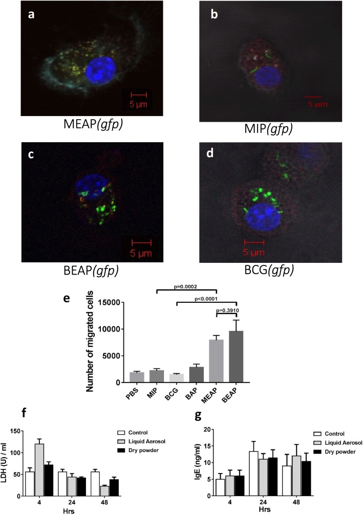
Fig. 3.
Fluorescent images showing the interaction between BMDCs and GFP-MIP, GFP-MIP encapsulated alginate micro-particles. a-d shows the co-localization of micro particles with the red stained lysosome. A representative cell after staining with different dyes was selected at random and pictures were taken in the four filter mode. The DCs after having phagocytosed MEAP(gfp) localized with lysosomes (red) appeared yellow (a). The CD11c was stained cyan and nucleus with DAPI (blue). b, c and d show MIP(gfp, BEAP(gfp) and BCG(gfp) respectively. e summarizes migration abilities of MEAP, BEAP, MIP, BCG and BAP activated BMDCs under the chemo-tactic influence of CCL21. 1 million BMDCs per well were plated in a 6 wells plate. They were treated with either 106 MIP/BCG or 50 μg MEAP/BEAP or 50 μg BAP suspended in PBS and incubated at 37 °C in 5% CO2 for 24 h. After 24 h, activated BMDCs were re-suspended in RPMI media and placed on the 5 μm pore sized transwell chamber. In the lower chamber of transwell plate, either PBS or CCL21 was placed. After 1 h, BMDCs migrated to the lower chamber were estimated by flowcytometry. f and g show comparison of LDH and IgE levels after delivering liquid aerosol and dry powder aerosol at different time points in the serum of mice

