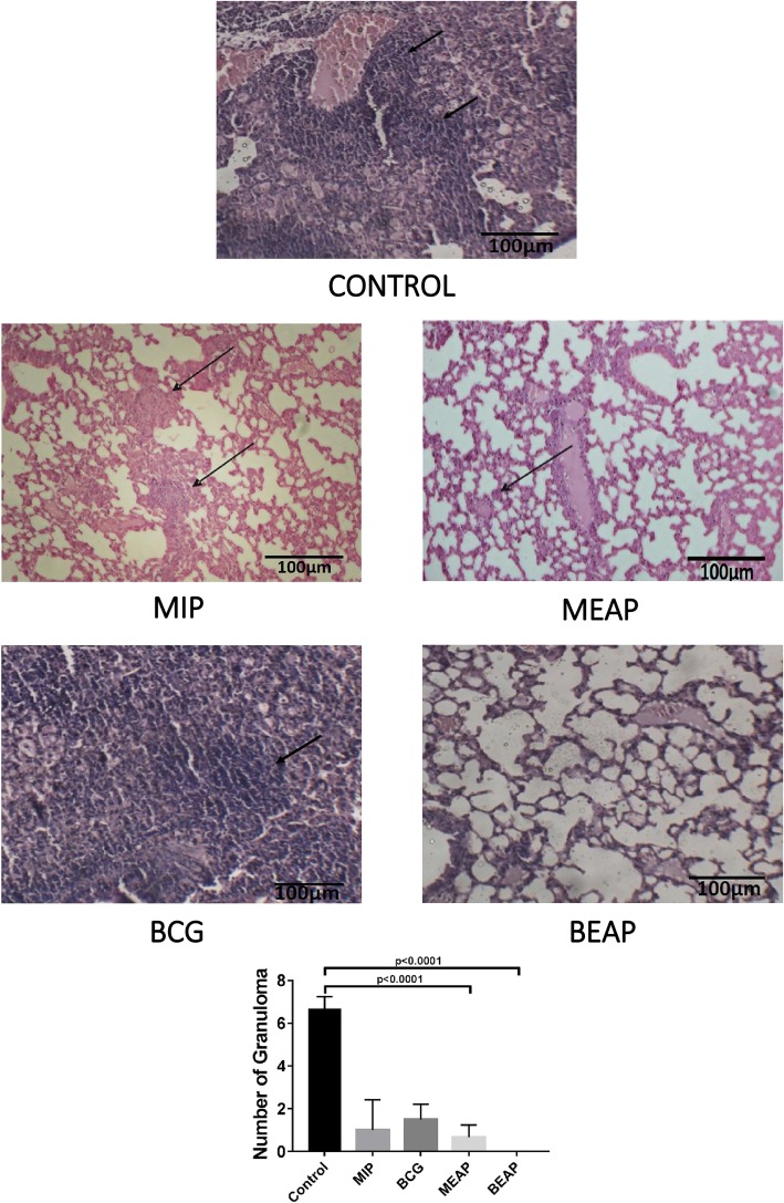
Fig. 6.
Histopathology of lungs from control, BCG, MIP, BEAP and MEAP immunized groups, post sixteen weeks of H37Rv infection. The arrows in figure indicate granulomatous lesions. The bar graph graph shows the summary of granulomatous lesions by randomly selecting 10 fields from 2 sections in each group

