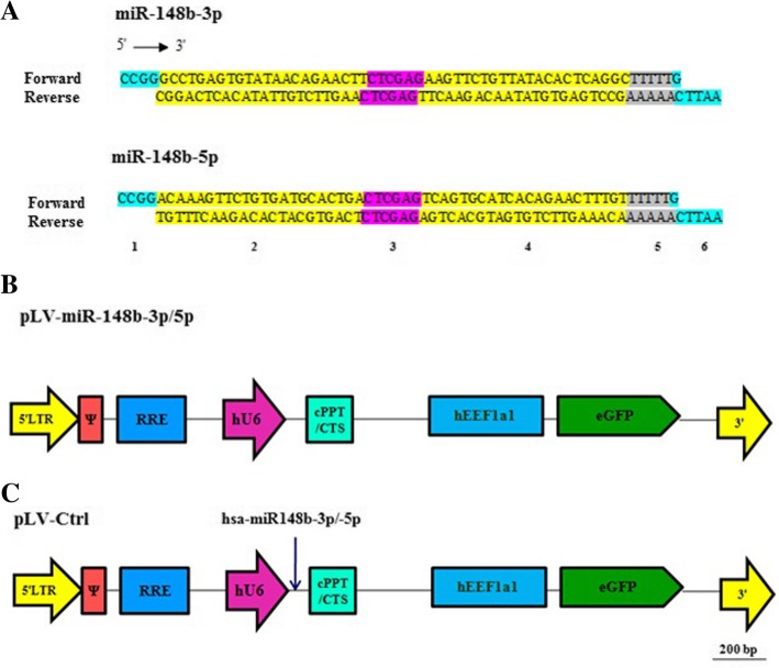
Fig. 1.
Cassettes and structure of LV shuttle plasmids. a Schematic representation of miR-148b-3p and miR-148b-5p sequences after ligation of forward and reverse primers. Section 1 and 6 are SgrA1 and EcoRI restriction sites, respectively. Section 2 and 4 are sense and antisense strands, respectively. Section 3 is necessary for hairpin structure (loop). Section 5 is a terminator (poly T). b Maps of pLV-miR-148b-3p and pLV-miR-148b-5p (c) and of pLV-Ctrl.5′ LTR, chimeric 5′ long terminal repeat including the Rous sarcoma virus U3 region as well as the HIV1 R and U5 regions; Ψ, HIV1 packaging signal; RRE, HIV1 Rev-responsive element; hU6, human U6 gene promoter; miR-158b-3p/5p, coding sequence of desired miRNAs; cPPT/CTS, HIV1 central polypurine tract and termination site; hEEF1a1, eukaryotic translation elongation factor 1 alpha 1 gene promoter; EGFP; enhanced green fluorescent coding sequence; 3′ LTR, 3′ HIV1 long terminal repeat involving a deletion in the U3 region to render the LV self-inactivating

