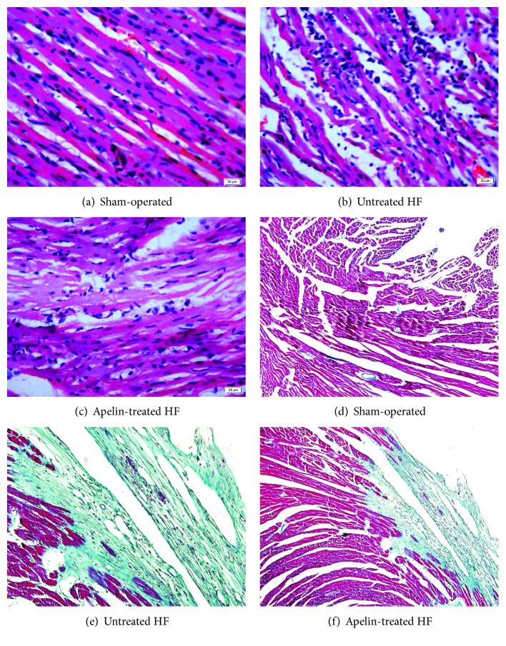
Figure 2.
The morphological changes as illustrated by hematoxylin and eosin staining and Masson's trichrome staining. For Masson's trichrome staining, fibrosis was stained blue, whereas cytoplasm red. Untreated HF animals displayed disorder structure and fibrotic myocardium, which were ameliorated by apelin treatment.

