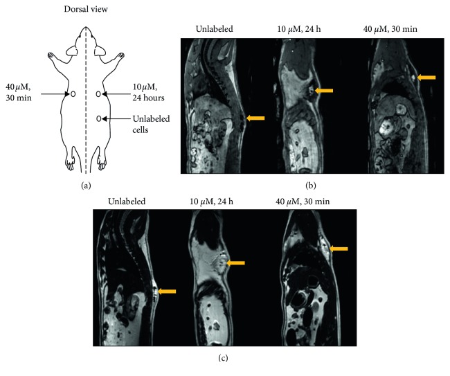
Figure 7.
In vivo MR imaging of transplanted hESCs in an adult rat. (a) Location of subcutaneous injections of hESCs in 0.2 mL mTeSR1 media on the dorsal side of rat. (b) 3D T 1-weighted TFE images without fat suppression clearly show contrast enhancement where the labeled cells were injected compared to unlabeled cells that were isointense against native tissue. (c) T 2-weighted TSE images were acquired to identify fluid present in all injections. Yellow arrows indicate location of injected cells.

