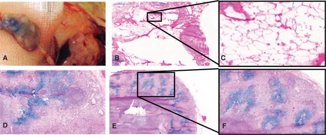
Figure 3:
Macroscopic and microscopic aspects of transplanted lymph node fragments (haematoxylin-eosin).
(A) Intraoperative macroscopic view. (B, C) Necrotic fragments presented fatty degeneration features observable within the lymph node capsule without major presence of lymphocytes. Vital fragments, in contrast, showed typical lymph node architecture (D–F) and sometimes germinal follicles (F). Here, blue-stained sinuses result from the peripheral injection of Berlin blue stain in the hind paws ante mortem. Scales: (B and F) 500 μm, (C) 100 μm, (D) 200 μm, and (E) 1000 μm.

