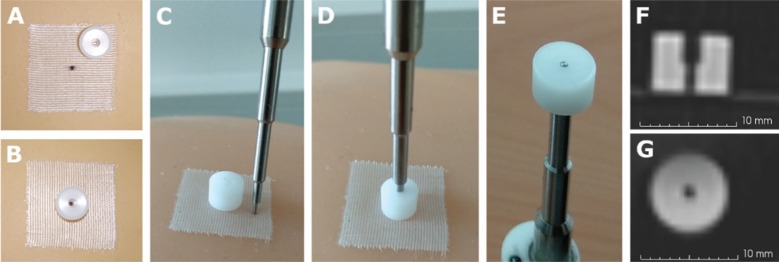
Figure 2:
External marker and pointer tip used for mapping the tracking and radiological data.
(A) External marker not aligned with the point marker. In this case, the point was drawn on the surgical tape. (B) External marker aligned with the point marker. (C) External marker and pointer tip. (D) Pointer tip inserted in the external marker. (E) Bottom view of the insertion. (F) CT slice with the lateral view of the external marker. (G) CT slice with the bottom view of the external marker. Voxel size 0.5×0.5×0.6 mm.

