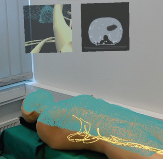
Figure 4:

Visualisation with HoloLens.
3D models of the phantom surface (blue wireframe) and vessels (yellow) superimposed on the FAST Ultrasound Training Model. 2D panels above the phantom show the point of view of the catheter (left, initial version of the Nav EVAR prototype) and an axial slice of the CT scan of the phantom (right). This photo was taken from the HoloLens user’s point of view after connecting to the web server Windows Device Portal on the HoloLens [38].
