
Figure 6:
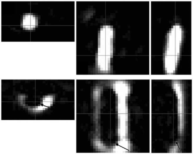
The axial, sagittal, and coronal images (left to right) show a balloon catheter labeled with MNPs (top) and a non-labeled balloon catheter in a vessel phantom filled with MNPs (bottom).
The FOV was 20×36×36 mm3 (with permission taken from Ref. [37]).