Figure 3.
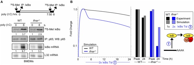
Type I interferon signaling potentiates late NFκB activity by translational inhibition of IκBα (A) Experiments to determine IκBα translation rate in BMDMs. Top, schematic of the experimental design: 35S-labeled Methionine pulsed at 0 h and following 8 h of Poly(I:C) stimulus. Middle, immunoprecipitates of IκBα following a 35S-methionine pulse at either indicated timepoint. NFkB p65 immunoprecipitates are shown as normalization controls. Bottom, IκBα mRNA analysis using RNA protection assay. Ribosomal protein gene L32 is provided as a control. These data are representative of three biological replicates. Quantitations are relative to basal conditions which is set to 1. (B) Using SiMoN to determine whether the measured changes in the translation rate are sufficient to account for the NFkB activation defect in ifnar−/− BMDMs. Left, timecourse simulation of NFκB activity in response to IKK activation following poly(I:C) stimulation with and without a 2-fold increase in IκBα translation measured in ifnar−/− BMDMs (A). Right, bar graph of NFκB activity at the peak and 24 h time point as quantified from the simulation and experiment (Figure 2A). This indicates that the increase in translation rate measured in (A) is not sufficient to account for the decrease in NFkB activity observed in Figure 2A.

