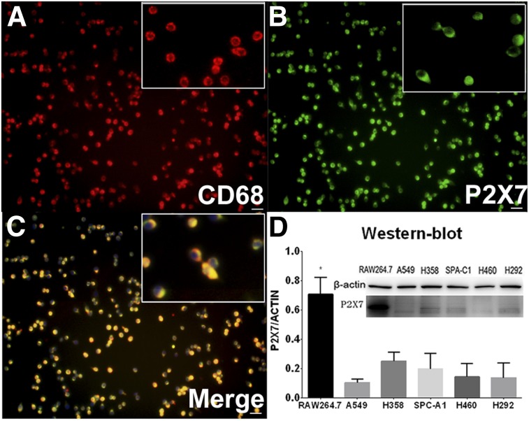
FIGURE 3.
Validation of P2X7R expression in cultured RAW264.7 cell line. (A–C) Immunofluorescence staining of CD68 (A), P2X7 (B), and merged (C) images showing marker localization in RAW264.7 cell culture. Images show that P2X7R and CD68 are expressed mainly on RAW264.7 cell membranes. Scale bars = 20 μm. (D) Western blot of RAW264.7 and series of lung cancer lines.

