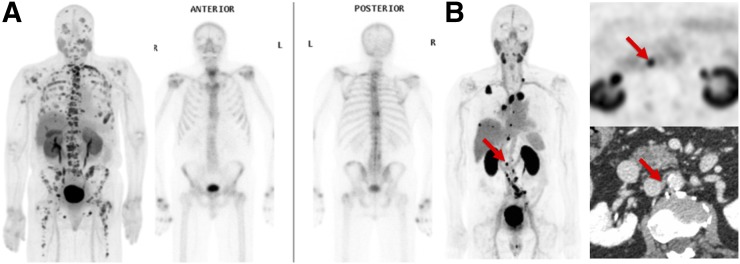
FIGURE 4.
(A) Example of CTT1057-avid osseous and lymph lesions from cohort B, showing CTT1057 PET maximum-intensity projection (left) and anterior (middle) and posterior (right) 99mTc-HDP planar bone scan. (B) Example of patient with extensive CTT1057-avid osseous metastatic disease and only minimal uptake on standard-of-care bone scan. CTT1057 MIP PET (left), axial PET (top right), and axial CT (bottom right) highlights patient with 4-mm short lymph node (arrows) that is not enlarged by size criteria on conventional CT but displays marked CTT1057 uptake indicative of high likelihood of metastatic involvement.

