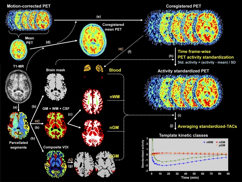
FIGURE 1.
Processing steps for acquiring 4 template kinetic classes. (a) By using FreeSurfer, T1-weighted MR images were segmented into 112 regions. (b) After merging regions in parcellated segments, masks for whole brain, gray and white matter, and composite VOI mask images were created. (c) White matter VOI masks least affected by activity of surrounding structures were created for each control. (d and e) PET images were coregistered to T1-MR image using mean PET image. (f) Sinus masks for each control were created. (g) In AD patients, masks for pathologic gray matter (pGM) were created. (h) Activity of PET images was standardized using whole brain masks. (i) Standardized time–activity curves for normal white matter (nWM), normal gray matter (nGM), and blood were obtained from 21 controls. Likewise, standardized time–activity curve for pGM was obtained from 25 AD patients. (j) By averaging standardized time–activity curves (TACs), template time–activity curves for 4 kinetic classes were established. HC = healthy control.

