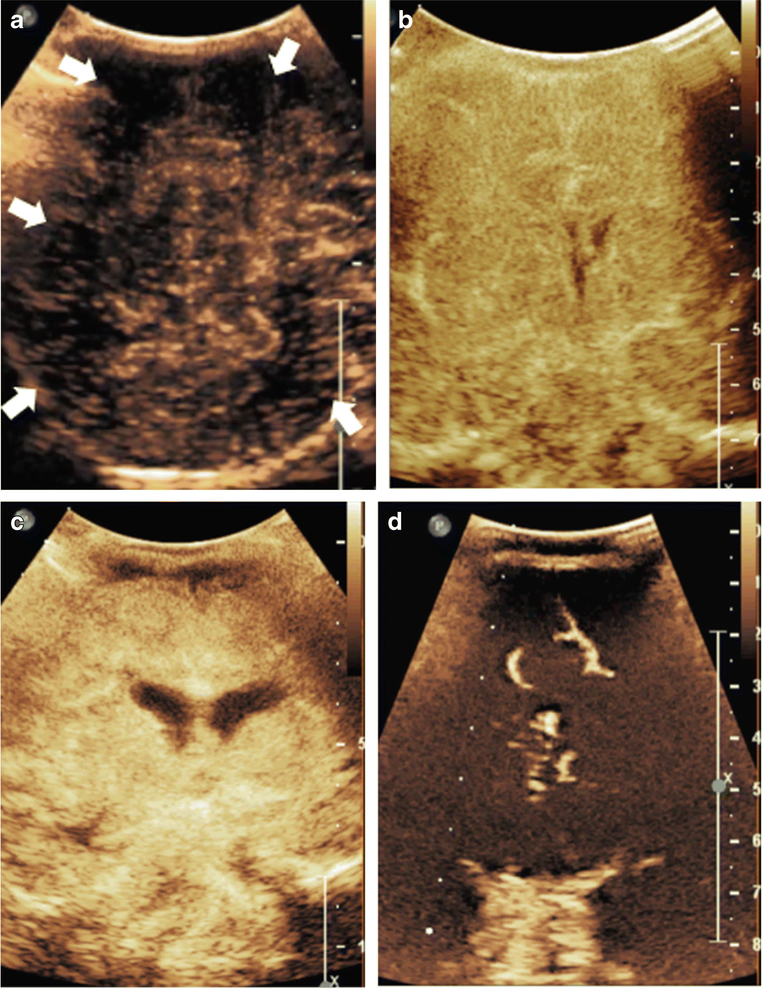
Figure 3. Multifocal and Symmetric, Diffuse Perfusion Abnormalities on CEUS-B.
Figure 3A depicts a coronal scan through the posterior parietooccipital lobes in a neonate with hypoxic ischemic injury. The image was obtained at 27 seconds after microbubble injection at peak intensity. Generalized hypoperfusion with multifocal perfusion abnormalities noted as evidenced by paucity of microbubbles in scattered areas (white arrows). Figure 3B and 3C, both obtained at 18 seconds after microbubble injection, are neonatal cases of symmetric, diffuse hypoxic ischemic injury resulting in generalized hyperperfusion to the brain in the immediate post-injury period. Figure 3D is an infant post prolonged cardiac arrest demonstrating diffuse hypoperfusion to the brain. Note that Figure 3 images were obtained with EPIQ scanner (Phillips Healthcare, Bothell, WA). Figure 3A was obtained with C5–1 transducer and settings of 12 Hz, MI 0.06, Figure 3B with C9–2 transducer and settings of 13 Hz, MI 0.06, Figure 3C with C9–2 transducer and settings of 7 Hz, MI 0.06, and Figure 3D with C9–2 transducer and settings of 12 Hz, MI 0.06. This figure is adapted from Hwang et al.[2] and Shin et al.[27] with permission.

