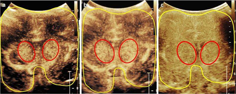
Figure 4. Central Gray Nuclei to Cortex Ratio (GNC).
Central gray nuclei to cortex ratio in a 1-month-old male (a, b) and a 14-day-old male (c). The central gray nuclei (basal ganglia and thalami) (red) and cortex (areas excluding central gray nuclei) (yellow) are outlined on a mid-coronal CEUS. The central gray nuclei to cortex (GNC) ratio equals perfusion in central gray nuclei (red) over perfusion in cortex (yellow). Figure (a) and(b) demonstrates a representative case with the normally seen hyperperfusion to the central gray nuclei, with (a) obtained at 10 seconds after microbubble administration and (b) obtained at 15 seconds after administration at peak enhancement. (c) demonstrates a hypoxic ischemic injury case in which the normally seen GNC ratio greater than 1 is absent due to diffuse hyperperfusion to the brain in the immediate post injury setting. In this case, the GNC ratio is noted to be approximately equal to 1. The image was obtained at 18 seconds after microbubble administration at peak enhancement. Note that all the images were obtained with EPIQ scanner (Philips Healthcare, Bothell, WA) and C5–1 transducer with settings of 12 Hz and mechanical index of 0.06.

