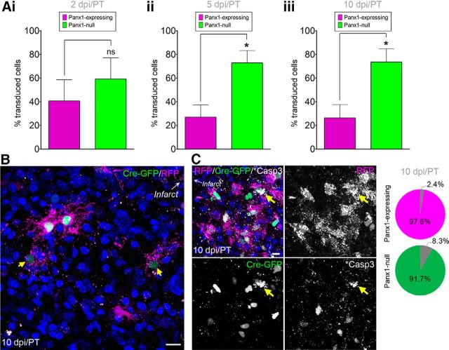
Figure 4.
Panx1-null NPCs persist in the peri-infarct cortex. A, Percentages of Panx1-null and Panx1-expressing NPCs in the peri-infarct cortex. Ai, Panx1-null and Panx1-expressing NPCs were equally abundant at 2 dpi/PT (n = 6, p = 0.4812 by unpaired t test). Note that 1 of the 7 brains did not have a single transduced NPC in the peri-infarct at 2 dpi/PT; data are represented as percentage of total transduced NPCs due to a large variability in NPC number reaching the peri-infarct cortex between mice. Aii, Panx1-null and Panx1-expressing NPC percentages at 5 dpi/PT (n = 4, p = 0.0208 by unpaired t test). Note that 2 of the 6 brains did not have a single transduced NPC in the peri-infarct at 5 dpi/PT. Aiii, Panx1-null NPCs were more abundant than Panx1-expressing NPCs at 10 dpi/PT (n = 6, p = 0.0180 by unpaired t test). B, Maximum-intensity projection of a representative confocal Z-stack from the peri-infarct tissue 10 dpi/PT. Arrows indicate faint GFP+ nuclei. C, Maximum-intensity projection of a representative confocal Z-stack from the peri-infarct tissue 10 dpi/PT with yellow arrows indicating activated caspase 3 (*Casp3)+ cells. Pie charts indicate the percentage of total RFP+ (Panx1-expressing; top) or GFP+ (Panx1-null; bottom) cells that were *Casp3+ in the peri-infact across all animals. Hoechst 33342 was used as a nuclear counterstain. Scale bars, 10 μm.

