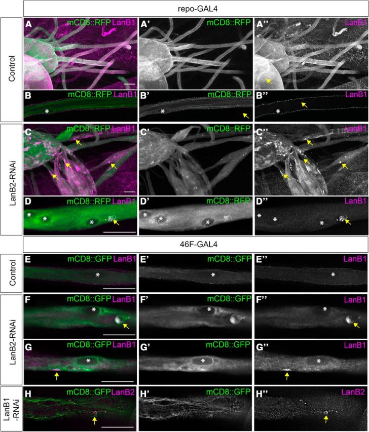
Figure 2.

LanB2 knockdown results in LanB1 accumulation in glia. A–D, Peripheral nerves of control (A, B) and repo>LanB2-RNAi larvae (C, D). Glia were labeled with mCD8::RFP (green) and immunolabeled for anti-LanB1 (magenta). A, C, Low-magnification images of peripheral nerves and ventral nerve cord. B, D, Higher-magnification images of individual peripheral nerves in longitudinal sections. Asterisks mark nuclei and arrows mark LanB1 accumulations. Scale bars, 30 μm. E–G, Longitudinal sections of peripheral nerves in control (E) and 46F>LanB2-RNAi larvae (F, G). Glia were labeled with mCD8::GFP (green) and immunolabeled with anti-LanB1 antibody (magenta). Asterisks mark nuclei and arrows mark LanB1 accumulations. Scale bars, 30 μm. H, Nerve from 46F>LanB1-RNAi larvae with glia labeled with mCD8::GFP (green) and immunolabeled with anti-LanB2 antibody (magenta). Arrows mark LanB2 accumulations. Scale bar, 30 μm.
