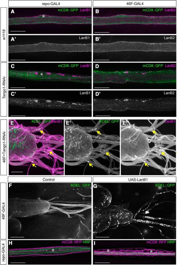
Figure 4.

Knockdown of Tango1 leads to accumulations of LanB1 and LanB2. A, B, High-magnification images of control repo-GAL4 (A) and 46F-GAL4 (B) peripheral nerves with glial membranes labeled with mCD8::GFP (green) and immunolabeled with LanB1 (A, magenta) or LanB2 (B, magenta). C, D, repo>Tango1-RNAi (C) and 46F>Tango1-RNAi (D) peripheral nerves demonstrate accumulations of LanB1 and LanB2 when Tango1 is knocked down (arrows). Nuclei are marked by asterisks. Scale bars, 20 μm. E, The ventral nerve cord and peripheral nerves of larvae where the perineurial glia driver 46F-GAL4 drove the expression of ER marker KDEL::GFP (green) and Tango1 RNAi. Colocalization of LanB1 accumulations (magenta) and ER aggregates was observed (yellow arrows). Scale bars, 50 μm. F, G, Low-magnification images of CNS and nerves with 46F-GAL4 driving KDEL::GFP without (F) or with (G) overexpression of LanB1 using UAS-LanB1EP-600, resulting in ER aggregates. Scale bars, 100 μm. H, High-magnification images of control nerves with glial membrane labeled with mCD8::RFP (magenta) and axons immunolabeled using anti-HRP antibody (green). I, Nerves with overexpression of LanB1 in glia, repo>LanB1EP-600, demonstrated vacuoles. Nuclei are marked by asterisks. Scale bars, 15 μm.
