
Figure 2.
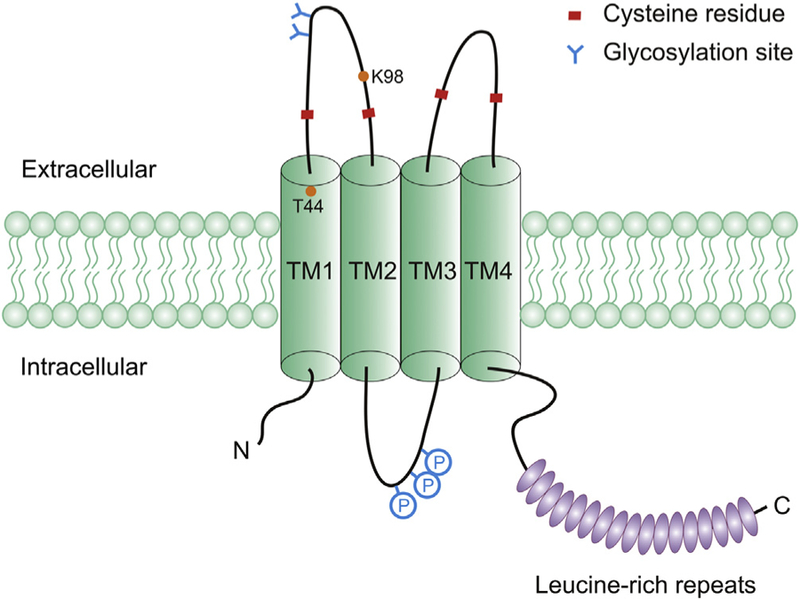
Schematic representation of SWELL1 protein. The diagram contains N-terminal 4 transmembrane helices (TM1–4), C-terminal 17 leucine-rich repeats (ovals), two pairs of conserved cysteine residues in the extracellular loops, and putative N-linked glycosylation sites and phosphorylation sites (P). Threonine (T44) located at the external boundary of TM1 and lysine (K98) within the TM1-TM2 extracellular loop were identifies as potential pore-lining residues.