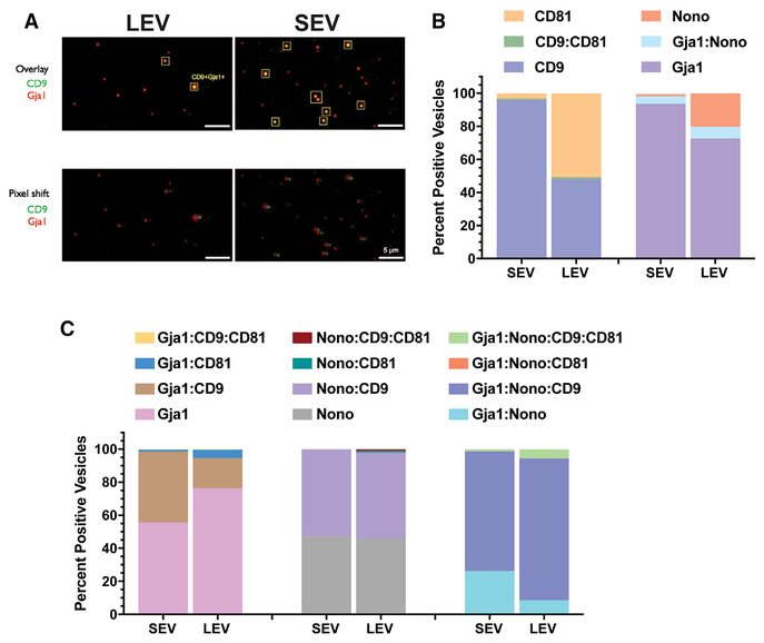
Figure 5. Single Vesicle Analysis of EV Fractions.
(A) Representative photomicrographs of immunofluorescence labeling of EV markers from large extracellular vesicles (LEVs) and small extracellular vesicles (SEVs) filtered fractions isolated from EGFR primary cultures. Pixel shift controls for co-registration of markers in overlay.
(B) Quantitative analysis (percentage) of singly or dually positive vesicles on a per vesicle basis for expression of CD9 and CD81 or Nono and Gja1 in LEV and SEV fractions.
(C) Quantitative analysis of singly, dually or triple combinations of indicated markers on a per vesicle basis for expression of CD9, CD81, Nono, and Gja1 in LEV and SEV fractions.
A total of 643 vesicles were analyzed from the LEV fraction and 1,648 vesicles were analyzed for the SEV fraction.
See also Figure S4.

