Figure 1.
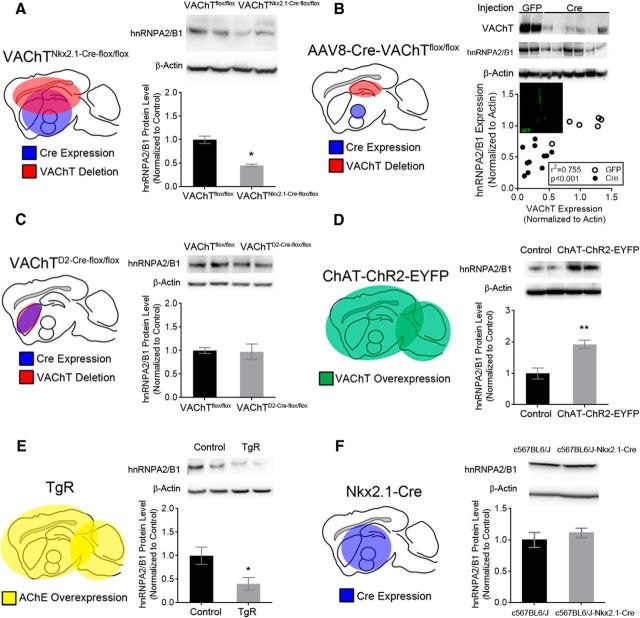
Analysis of hnRNPA2/B1 protein levels in genetically modified mice with differential expression of VAChT. A, Representative Western blot and quantification of hnRNPA2/B1 protein expression in the hippocampus of VAChTflox/flox and VAChTNkx2.1-Cre-flox/flox mice. hnRNPA2/B1 expression was normalized to actin (n = 6). B, hnRNPA2/B1 protein levels positively correlate with AAV induced reduction of VAChT. The graph shows values for each individual mice injected either with GFP or CRE. The image is from the medial septum of a virus-injected mice. C, Striatal elimination of VAChT does not alter hnRNPA2/B1 protein levels in the striatum of VAChTD2-Cre-flox/flox mice (n = 4). D, Transgenic mice overexpressing VAChT have increased hnRNPA2/B1 protein levels compared to controls (n = 4). E, hnRNPA2/B1 protein levels from the hippocampus of TgR transgenic mice (n = 3). F, No change in hnRNPA2/B1 protein levels compared to controls in the hippocampus of c567BL/6J-Nkx2.1-Cre mice (n = 3). Data are mean ± SEM. *p < 0.05; **p < 0.01.

