Figure 3.
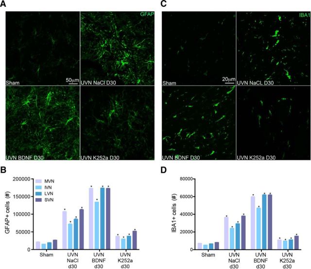
BDNF treatment increases astrocytic and microglial cell population in the VN. A, C, Illustrations of GFAP− (A) or IBA1+ (C) cells in the left (lesioned) MVN of sham, UVN-NaCl, UVN-BDNF, and UVN-K252a animals 30 d after UVN. Scale bars: A, 50 μm; B, 20 μm. n = 4 animals per group. B, D, Histograms showing the effects of NaCl, BDNF, or K252a continuous infusion on the number of GFAP− (B) or IBA1+ (D) cells in the four VN 30 d after UVN. Only values recorded on the lesioned side are illustrated and data from both sides of the sham group were pooled for direct comparison with the subgroups of vestibular deafferented cats. SEMs are shown as vertical lines. Analyzes were assessed by ANOVA followed by Scheffe test for all of the VN and all groups (*p < 0.0001 vs the same VN of other groups).

