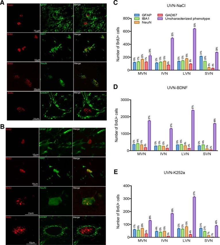
Figure 5.

BDNF influences the cell differentiation pattern of newborn cells in the deafferented VN. Confocal analysis of differentiated newly generated cells by double immunostainings processed on consecutive serial sections in the deafferented MVN of cats infused with BDNF (A) or K252a (B) infusion from days 0–30 after UVN. The BrdU+ nuclei are shown in red and the other markers of differentiation (GFAP, IBA1, NeuN, and GAD67) are shown in green. Histograms illustrating the percentage are defined as the ratio between the mean number of immunopostive elements colocalizing a cell-type marker (GFAP, IBA1, NeuN, or GAD 67) and BrdU relative to the total mean number of BrdU+ nuclei counted in the areas of quantification of the UVN-NaCl (C), UVN-BDNF (D), or UVN-K252a (E) groups. SEMs are shown as vertical lines.
