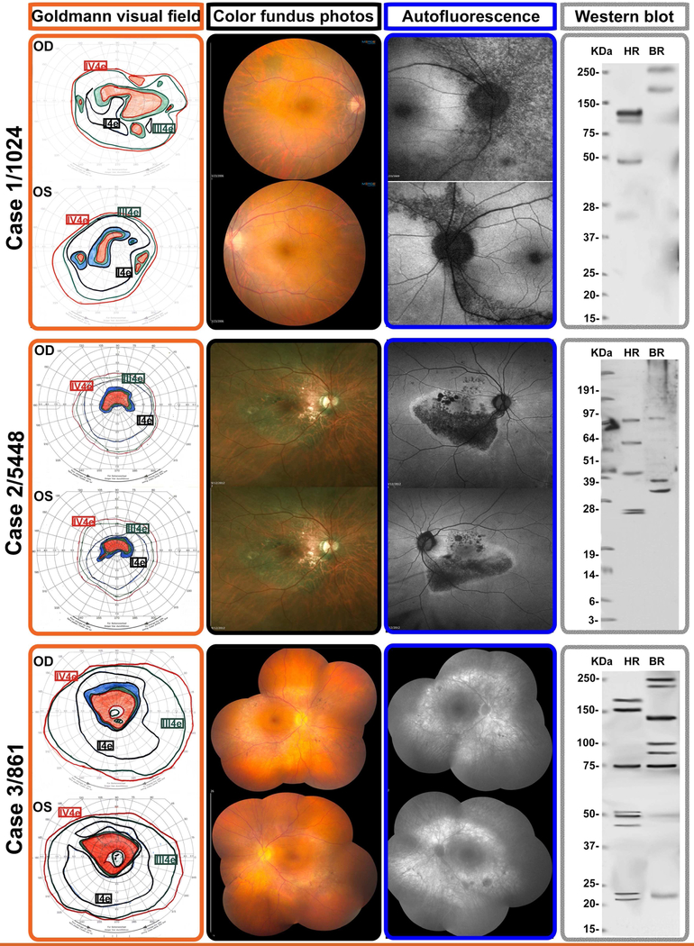
Figure 1-. Representative multimodal imaging of 3 patients presenting with Acute zonal occult outer retinopathy.
Selected cases to illustrate the testing and imaging findings of 3 patients presenting with typical AZOOR findings as defined by the criteria originally defined by Gass et al. These correspond to patients listed in Table 1 and identified with their laboratory identification number
Top row: Case 1 is patient 1024, a 42-year-old woman who presented with progressive two-and-a-half year history of decreased visual acuity and distortion of fine print, OD worse than OS. The Goldmann visual fields showed enlarged blind spots and peripheral scotomata OS>OD. Fundus examination showed no overt lesions and mild vascular attenuation; the autofluorescence (AF) demonstrated RPE damage emanating from the discs OU. Western blot showed five anti-retinal antibody bands in the human test lane.
Middle row: Case 2 is patient 5448, a 58-year-old man who presented with a two-year history of bilateral peripheral visual field loss and central vision blurring. He had a family history of diabetes and connective tissue disease. His autofluoresence was remarkable for RPE damage emanating from focal points of the optic disc, which correspond exquisitely well to the color fundus photographs and the outline of his GVF. He had four anti-retinal antibody bands on Western blot.
Bottom row: Case 3 is patient 861, a 63-year-old lady with a one-year history of visual distortion OS>OD. Goldmann visual field testing showed full isopters, but with pericentral scotomatous regions OU, consistent with the areas of atrophy in her fundus. Fundus color photographs and autofluorescence show areas of diffuse RPE loss corresponding to the visual field scotomata. The pattern of RPE damage OU was suggestive that toxic immune products leaked from the disc margins and spread into the subretinal space. There were minimal pigmentary deposits in the areas of RPE atrophy. She had eight anti-retinal immunoreactive bands on Western blot

