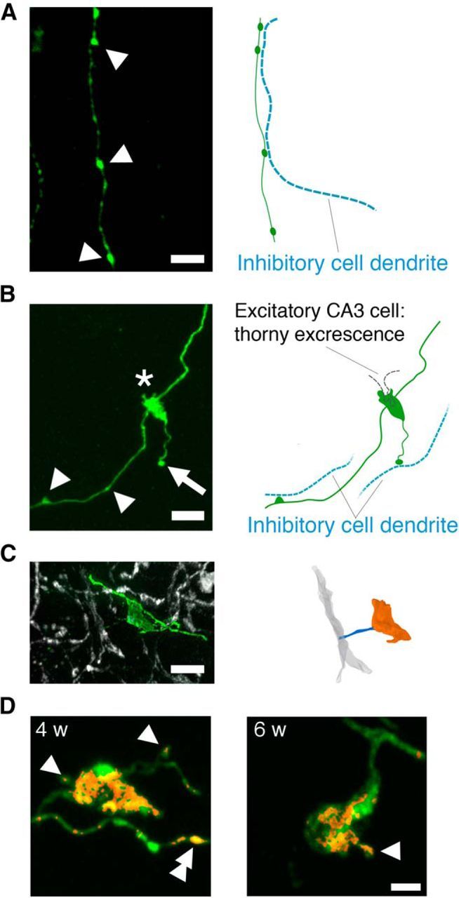
Figure 2.

High-resolution images of the three major mossy fiber presynaptic specializations. A, Left, Retrovirally labeled mossy fiber in the hilus. En passant boutons (arrowheads) form contacts with dendrites of inhibitory cells within the hilus. Scale bar, 5 μm. Right, Putative location of dendrites of inhibitory cells within the hilus is shown as blue dotted lines. B, Retrovirally labeled cells extend their mossy fiber axon to the CA3-sl region and develop specialized presynaptic contacts with principal excitatory CA3 pyramidal cells via LMTs (asterisk) or local inhibitory CA3 interneurons via en passant boutons (arrowheads) or filopodia extensions from LMTs (arrow). Scale bar, 5 μm. Putative locations of dendrites/thorny excrescence of principal excitatory CA3 pyramidal cells (black dotted line) and inhibitory CA3 interneurons (blue dotted line) are depicted in the right. C, Representative GFP+ filopodia (green) extending from an LMT making contact with a parvalbumin+ dendrite (gray) of an inhibitory CA3 neuron. Scale bar, 5 μm. D, Representative GFP+ LMTs and associated filopodia (green) 4 and 6 weeks after infection. Bassoon (red) is expressed in LMTs, the head of filopodia (single arrowhead), and en passant boutons (double arrowhead). The Bassoon channel was masked using the GFP channel. Scale bar, 2 μm.
