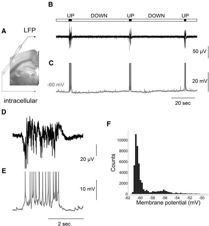
Figure 1.
Spontaneous synchronized network activity in the form of Up/Down states recorded in a mouse brain slice. A, Picture of a coronal slice used for simultaneous LFP (black electrode) and intracellular (gray electrode) recording from layer 2/3 of the primary somatosensory cortex. B, C, LFP and intracellular recordings, respectively. The bar above the traces indicates Up states (in black) and Down states (in gray). Action potentials are truncated at −30 mV. D, E, Close-up of the LFP and intracellular recording, respectively, showing an Up state at higher resolution. F, Membrane potential distribution with two peaks reflecting the bistable fluctuations of membrane potential during spontaneous network activity.

