
Figure 6.
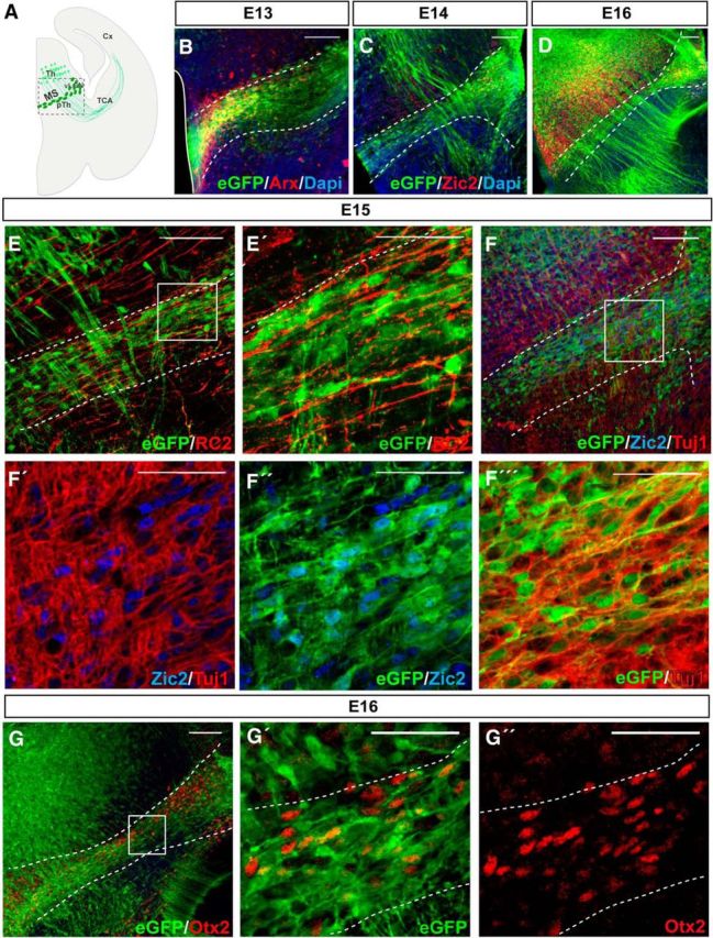
Zic2 is expressed in thalamus–prethalamus boundary INs. A, Scheme representing a coronal section from an E13.5 mouse brain. The squared area delimits the zone depicted in all the images of this figure. B, Arx immunostaining in coronal sections of E13.5 Tg(Zic2eGFP) embryos confirm the existence of a population of Zic2/eGFP cells in the thalamic-prethalamic boundary. White dashed lines delimit the thalamic prethalamic boundary (TpTB) Zic2-positive area. C, D, Zic2/eGFP cells in the thalamic–prethalamic boundary at E15.5 and E16.5. Note that Zic2 is also expressed in the thalamus and that thalamocortical axons are also positive for eGFP and visible from E14.5 in advance. E, E′, Immunostaining for RC2 in coronal sections of E15.5 Tg(Zic2eGFP) embryos demonstrate that Zic2/eGFP cells are not glial cells. F–F′′′, Zic2 and Tuj1 immunostaining in coronal sections of E15.5 Tg(Zic2eGFP) embryos demonstrate that Zic2/eGFP cells in the TpTB are neurons. Note that eGFP cells present Zic2 staining in the nucleus. G–G′′, Otx2 immunostaining in coronal sections of E15.5 Tg(Zic2eGFP) embryos demonstrate that Zic2/eGFP cells are INs. Th, thalamus; pTh, prethalamus, Cx, cortex; TCA, thalamocortical axons. Scale bars: B–G, 100 μm; E′–G′′, 50 μm.