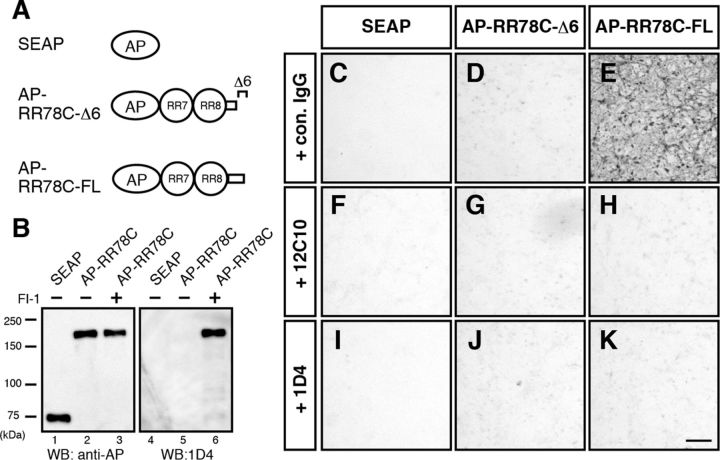
Figure 5.
RR78 with intact CTR binds to neuronal cell membrane. A, Schematic of the secreted AP (SEAP), AP-RR78C protein collected in the presence of DMSO (as a vehicle, AP-RR78C-Δ6), and AP-RR78C collected in the presence of FI-1 (AP-RR78C-FL). B, SEAP or AP-RR78C was expressed in HEK293T cells in the presence of DMSO or 50 μm FI-1. Culture supernatants were analyzed by WB using anti-AP and 1D4 antibodies. Molecular mass markers (kDa) are shown on the left of the panel. C–K, AP staining of primary cultured cortical neurons. Primary cultured cerebral cortical neurons were incubated with SEAP (C, F, I), AP-RR78C-Δ6 (D, G, J), or AP-RR78C-FL (E, H, K) in the presence of anti-myc antibody as the control IgG (C–E), 12C10 (F–H), or 1D4 (I–K). The interaction between AP-RR78C-FL and the neuronal cell membrane was inhibited by 12C10 or 1D4 antibodies (H and K, respectively). Scale bar, 100 μm.

