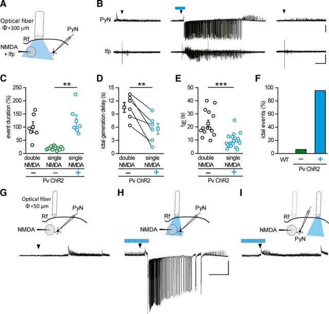
Figure 3.
Optogenetic activation of Pv interneurons at the epileptogenic focus promotes ictal event generation. A, Schematic of the experiment and (B) representative voltage-clamp recording (Vh = −50 mV) from a pyramidal neuron 700 μm from the focus and simultaneous local field potential recording in response to a single NMDA pulse in the absence (left, right) and in the presence (middle) of light stimulation at 0.5 Hz, which was initiated 30 s before the NMDA challenge. Note the ictal response evoked by a single NMDA pulse in the presence of optogenetic stimulation. Calibration: 40 s, 0.01 mV, 500 pA. C, Distribution of normalized event duration in double NMDA pulses (black circles, n = 6, 4 mice) and single NMDA pulse experiments in the absence (green circles, n = 12, 4 mice) and in the presence (cyan, n = 6, 4 mice) of light stimulation. As in the representative experiment reported in B, in the six experiments the effect of a single NMDA pulse was checked twice, both before and after the ictal event evoked by the single NMDA pulse coupled with light stimulation. All data points are normalized to the mean duration of double NMDA-evoked events. D, Paired distribution of the delay in ictal onset measured from local field potentials recorded at the focus in a subset of the experiments (n = 6 ictals, 4 mice) that are reported in E. E, Distribution of the delay between the first NMDA pulse and the recruitment of the pyramidal neuron, as measured by tIE (see Materials and Methods), in ictal events evoked by double NMDA pulses in the absence of light stimulation (black, n = 14, 7 mice), and single NMDA pulses in the presence of light stimulation (cyan, n = 20, 7 mice). F, Bar histogram of the percentage of ictal events evoked by a single NMDA pulse in slices from Pv-Cre mice injected with saline solution (n = 9 trials, 3 mice) and from ChR2 Pv mice in the absence (−, green bar, n = 33 trials, 9 mice) and in the presence (+, cyan, n = 23 trials, 9 mice) of pulsed light stimulation. G–I, Schematic of the experiment (top) and representative voltage-clamp (Vh = −50 mV) recordings (bottom) of the response to a single NMDA pulse in a pyramidal neuron located 250 μm from the focus in the absence of light stimulation (G), in the presence of light stimulation at the focus (H), and in the presence of light stimulation in distant regions 1 mm from the focus (I) (n = 3 ictal events, 2 mice). **p < 0.01. ***p < 0.001.

