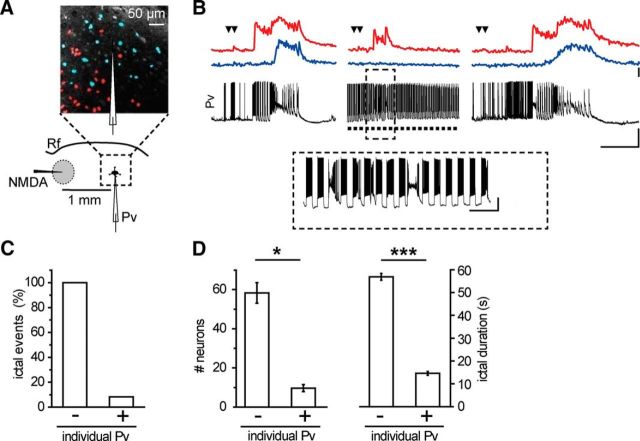
Figure 8.
Individual Pv interneuron activation in penumbra regions blocks ictal event propagation. A, Schematic of the experiment and differential interference contrast image illustrating the neuronal clusters progressively recruited into the ictal event in Ca2+ imaging experiments. B, Representative single current-clamp recordings from a Pv interneuron and simultaneous mean Ca2+ signal from two neuronal clusters (red and blue traces; soma position of these neurons is reported in A according to their different time of recruitment into the propagating ictal event. Steps of current were repeatedly injected to evoke firing activity (inset) that prevented the occurrence of a full ictal event. Calibration: 40 s, ΔF/F0 40%, 40 mV; inset, 5 s, 20 mV. C, Bar histogram of the percentage of NMDA evoked ictal events without (−, n = 23, 8 mice) or during (+, n = 12, 8 mice) stimulation of individual Pv interneurons. D, Bar histograms reporting the number of recruited neurons (left) and ictal event duration (right) measured in red neuron Ca2+ signals, in the absence and in the presence of current pulse stimulation of Pv interneurons (n = 3 ictal events, 2 mice). *p < 0.05. ***p < 0.001.

