
Figure 2.
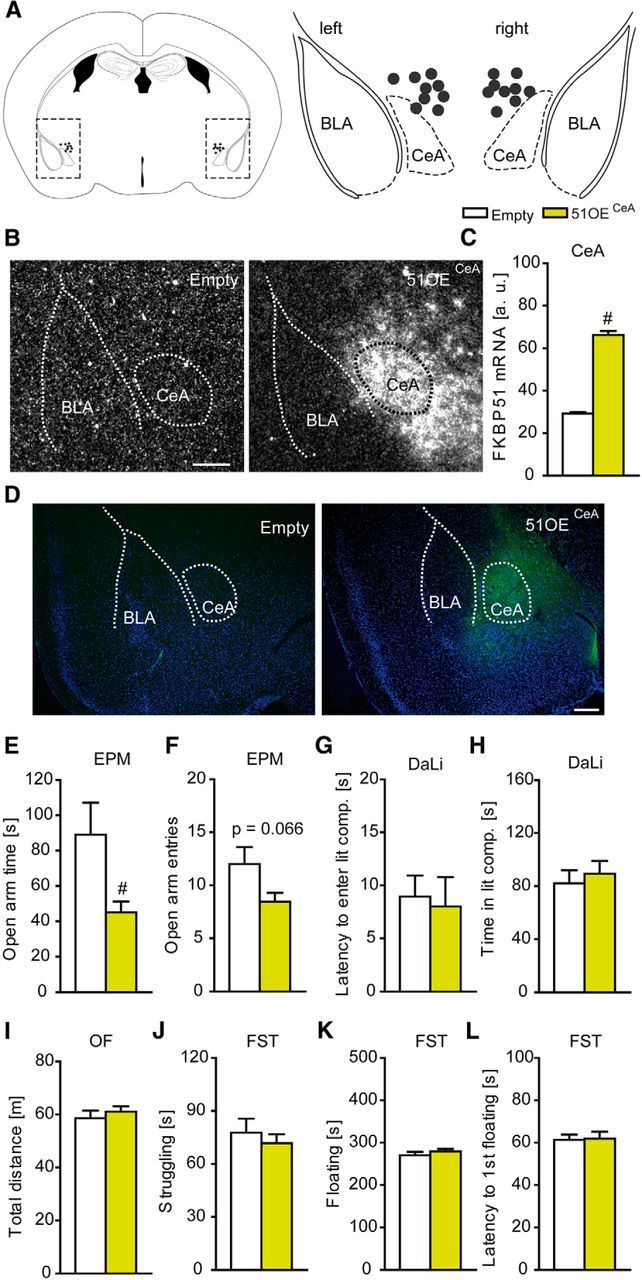
Overexpression of FKBP51 in the CeA. A, Schematic representation of the viral injection into the CeA. Gray dots represent the location of the injection tip. B, Representative autoradiographs of viral FKBP51 mRNA expression in the CeA 7 weeks after injection .C, Quantification of FKBP51 mRNA levels in the CeA. Infection with the viral construct induced a strong increase. D, Representative immunohistochemistry images of viral FKBP51 expression (Ha-tag Immuno, green; DAPI, blue) in the CeA region 7 weeks after injection. E, F, FKBP51 overexpression in the CeA leads to significantly reduced open arm time and a trend toward decreased open arm entries in the EPM. G, H, 51OECeA mice show no alterations in the DaLi. I–L, General locomotion as assessed in the OF and stress-coping behavior in the FST are not affected by FKBP51 overexpression in the CeA; n = 9–10; #p < 0.05. Data are expressed as mean + SEM. Scale bars, 250 μm.