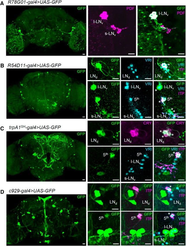
Figure 2.

Characterization of gal4 strains. gal4 lines were crossed with uas–GFP–S65t to visualize the gal4 expression patterns. Flies were entrained to 12/12 h LD for 5 d and were dissected at ZT 19. Brains were triple stained with anti-GFP (green), anti-VRI (cyan), and anti-PDF (magenta in A) or anti-ITP (magenta in bottom rows of B–D). The ITP-positive LNd and fifth s-LNv are CRY-positive neurons (Johard et al., 2009). trpA1SH–gal4/uas–GFP flies were double stained with anti-GFP (green) and anti-dCRY (magenta) to determine whether GFP-positive LNd were also CRY positive (C, top row). Scale bars, 10 μm.
