
Figure 7.
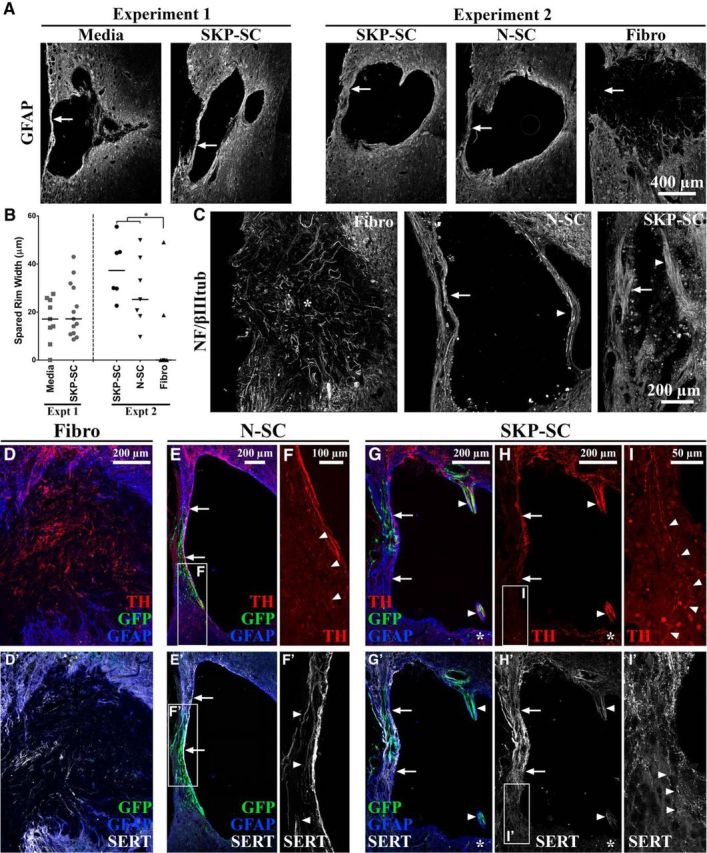
Relative to fibroblasts, transplanted SCs enhance the substrate available for axons to traverse the lesion site from rostral to caudal. A, Representative images (based on group median spared rim widths) of GFAP-immunolabeled tissue at the level of injury depicting the relative sizes of the spared tissue rims (arrows) in each treatment group. B, Spared rim width (μm) depicted as individual data points with group medians indicated by solid black lines (*p < 0.0167, Bonferroni-adjusted significance level for Experiment 2 pairwise tests). Both SC groups had significantly thicker spared rims than the Fibro group (Experiment 2) becaue the majority of animals in the latter group had no spared rim at the lesion epicenter (A, B). C, Sample images of NF/βIIItub+ axons in each group in Experiment 2. Axons were oriented rostro–caudal in the spared rim (arrows) and midlesion bridges (arrowheads) of N-SC- and SKP-SC-treated tissue, whereas axons within the lesion site of animals in the Fibro group were oriented toward the middle of the lesion (asterisk in C) and/or the lateral edge of the spinal cord (i.e., the periphery). D–I′, Sample images from animals in each group in Experiment 2 immunolabeled for GFP (green), GFAP (blue), and TH (red; D–H, I) or SERT (white; D′, E′, F′, G′, H′, I′). Similar to NF/βIIItub+ axons (C), TH+ and SERT+ axon subpopulations both lacked appropriate rostral-caudal orientation in the Fibro group (D, D′), but crossed the spared tissue rim (arrows in E, E′ [N-SC] and G, H, G′, H′ [SKP-SC]) or midlesion bridges (arrowheads in G, H, G′, H′) in the SC groups. In the latter, axons were found in intact GFAP+ regions or regions occupied by GFP+ SCs (arrowheads in F′ [higher magnification image of the box in E′]). The density of TH+ and SERT+ axons was highest rostral to the lesion and lowest caudal to the lesion in both SKP-SC (G, G′) and N-SC (E, E′) groups and at the caudal extent of the lesion TH+ and SERT+ axons appeared to exit the spared rim (arrowheads in F, I, I′ [higher-magnification images of the boxes in E, H, H′]) and the midlesion bridges (astrisks in G, H, G′, H′). All data are from longitudinal tissue sections at 11 wpi.