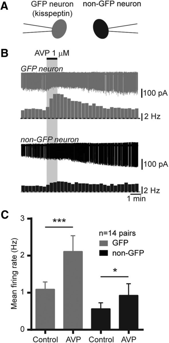
Figure 4.

AVP activates kisspeptin and other neurons in the RP3V. A, Diagram illustrating the recording configuration. GFP (gray) and non-GFP (black) neurons were recorded simultaneously. B, Example paired recording and corresponding rate meters (20 s bins) showing the excitatory effect of AVP (1 μm) in an RP3V kisspeptin neuron (gray) and a non-GFP RP3V neuron (black). C, Summary bar graphs of the effect of AVP in kisspeptin neurons and in unidentified neurons in the RP3V. *p < 0.05, Student's t test; ***p < 0.001, Wilcoxon matched pairs test.
