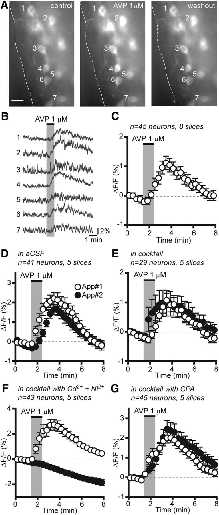
Figure 6.

AVP increases [Ca2+]i in RP3V kisspeptin neurons. A, Average projection images (1 min) of an RP3V slice before, immediately after, and 5 min after the application of AVP (1 μm). Scale bar, 10 μm. B, Corresponding traces showing that AVP reversibly increased fluorescence in multiple RP3V kisspeptin neurons. C, Average time course of GCaMP3 fluorescence changes in response to AVP in RP3V kisspeptin neurons from diestrous mice. D–G, Average time course of RP3V kisspeptin neuron [Ca2+]i responses to two successive applications of AVP (App#1 and App#2) in aCSF (D), in the presence of a cocktail including a sodium channel inhibitor, and GABAA, AMPA, and NMDA receptor antagonists (E), in the cocktail supplemented with Cd2+ and Ni2+ (F) and in the cocktail supplemented with CPA (G; cocktail contained TTX, CNQX, AP5, and gabazine). Each data point represents the relative GCaMP3 fluorescence (20 s bins) averaged across multiple neurons in C–G.
