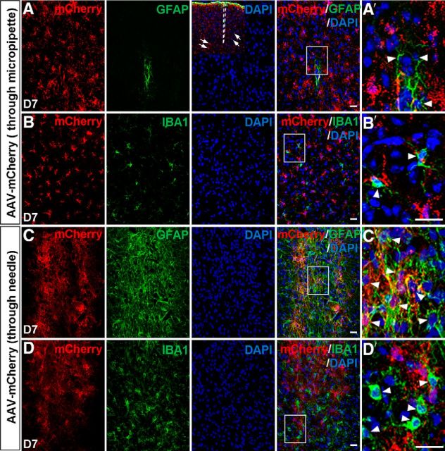
Figure 10.
Injured states induced by micropipette or needle injections in the dorsal midbrain. A, B, Immunostaining of mCherry, GFAP (A), or IBA1 (B) on sections of the dorsal midbrain from adult mice that were infected with AAV–mCherry through micropipettes (18–20 μm in diameter) at 7 DPI. The inset in A shows that cells (arrows) away from the injection site were analyzed. A′ and B′ are higher magnification of the boxed areas in A and B, respectively. C, D, Immunostaining of mCherry, GFAP (C), or IBA1 (D) on sections of the dorsal midbrain from adult mice that were infected with AAV–mCherry by 31 gauge needles (∼260 μm in diameter) at 7 DPI. C′ and D′ are higher magnification of the boxed areas in C and D, respectively. Scale bars, 20 μm.

