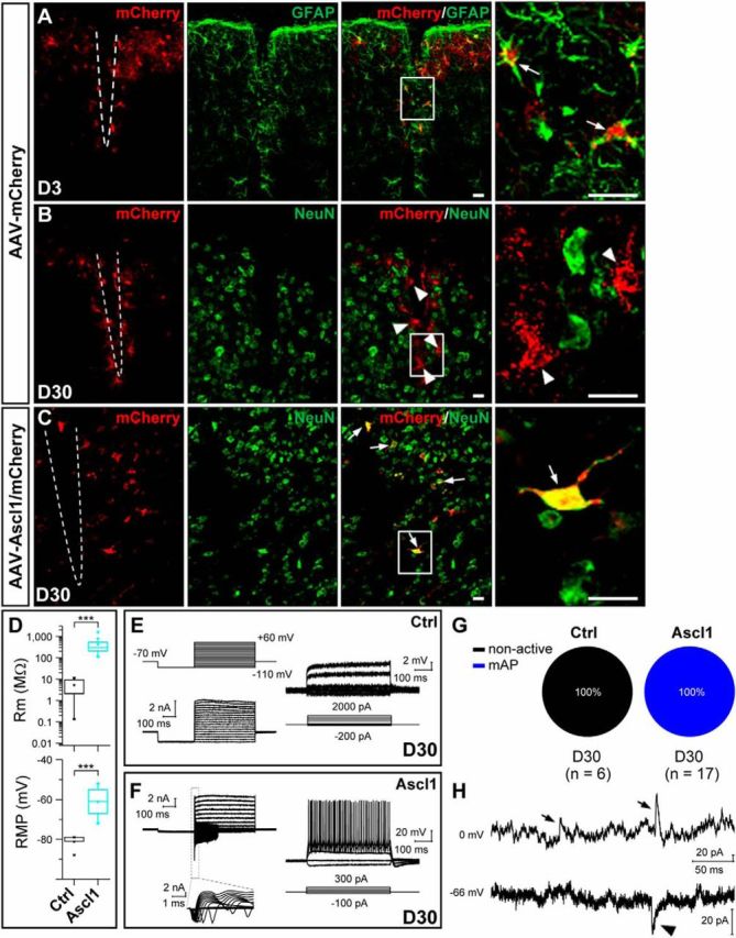
Figure 11.

Conversion of reactive astrocytes into neurons by Ascl1. A, Double staining of mCherry and GFAP on sections of the injured dorsal midbrain from adult mice that were infected with the control virus AAV–mCherry (A) at 3 DPI. Most mCherry+ cells expressed GFAP in the injured site. B, C, Double staining of mCherry and NeuN on sections of the injured dorsal midbrain from adult mice that were infected with the control AAV–mCherry (B) or AAV–Ascl1/mCherry (C) virus at 30 DPI. D, Rm and RMP of induced cells of the injured dorsal midbrain in mice 30 d after infection of the control AAV–mCherry or AAV–Ascl1/mCherry virus. E, F, Membrane currents (left) and voltages (right), elicited by step voltage and current commands, respectively, of an example mCherry+ cell in the slice of injured dorsal midbrain from a WT mouse 30 d after infection of the control virus AAV–mCherry (E) or an example iN cell (mCherry+) in the slice of dorsal midbrain from a WT mouse 30 d after the infection of the AAV—Ascl1/mCherry virus (F). G, Percentages of induced cells with different degrees of membrane excitability (non-active and mAP) in mice 30 d after infection of the control AAV–mCherry or AAV–Ascl1/mCherry virus. H, Representative spontaneous inward (putative glutamatergic, arrowhead) and outward (putative GABAergic, arrow) synaptic currents recorded at Vclamp = −66 and 0 mV, respectively, from an iN cell at 30 DPI. Scale bars, 20 μm. Ctrl, Control. Kolmogorov–Smirnov test was used in D. ***p < 0.001. Error bars indicate SEM.
