Figure 2.

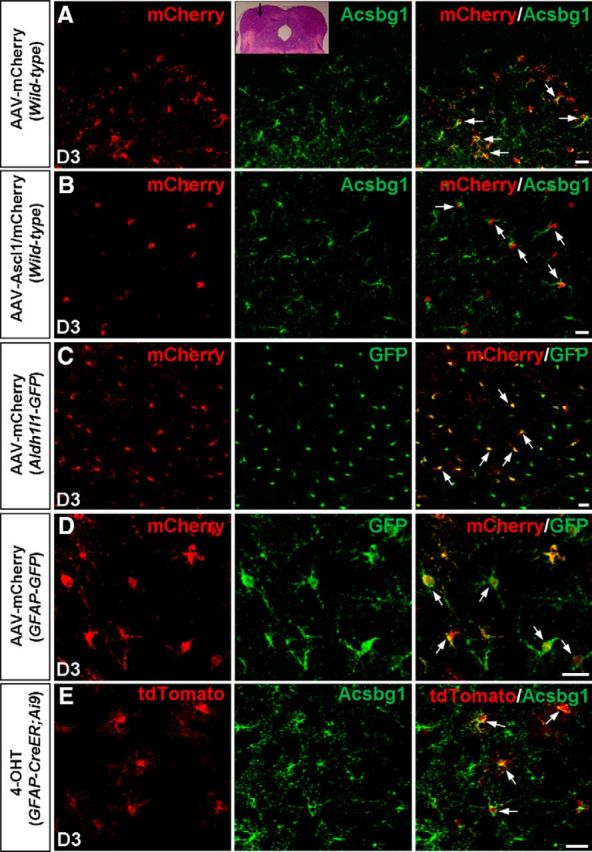
GFAP–AAV vectors target astrocytes of the dorsal midbrain in vivo. A, B, Double staining of mCherry and Acsbg1 on sections of the dorsal midbrain from WT mice infected with the control virus AAV–mCherry (A) or virus AAV–Ascl1/mCherry (B) at 3 DPI. mCherry was almost expressed exclusively in astrocytes. The inset shows the area where the AAV viruses were injected. C, D, Double staining of mCherry and GFP on sections of the dorsal midbrain from Aldh1l1–GFP (C) and GFAP–GFP (D) mice infected with AAV–mCherry at 3 DPI. mCherry extensively colabeled with GFP (arrows). E, Double staining of tdTomato and Acsbg1 on sections of the dorsal midbrain from GFAP–CreERT2;Ai9 mice that were injected with 4-OHT. tdTomato extensively colabeled with Acsbg1 (arrows). Scale bars, 20 μm.
