
Figure 5.
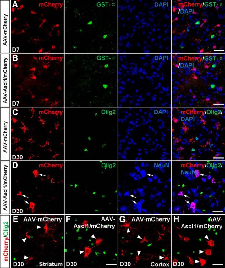
Ascl1 converts astrocytes to neurons but not oligodendrocytes. A, B, Double staining of mCherry and GST-π on sections of the dorsal midbrain from WT mice infected with the control virus AAV–mCherry (A) or virus AAV–Ascl1/mCherry (B) at 7 DPI. mCherry was not colocalized with GST-π. C, Double staining of mCherry and Olig2 on sections of the dorsal midbrain from WT mice infected with the control virus AAV–mCherry (C) at 30 DPI. mCherry was not colocalized with Olig2. D, Triple staining of mCherry, Olig2, and NeuN on sections of the dorsal midbrain that were infected with AAV–Ascl1/mCherry at 30 DPI (D). mCherry was colocalized with NeuN but not with Olig2 (arrows). E, F, Double staining of mCherry and Olig2 on sections of the striatum from WT mice infected with the control virus AAV–mCherry (E) or virus AAV–Ascl1/mCherry (F) at 30 DPI. G, H, Double staining of mCherry and Olig2 on sections of the cortex from WT mice infected with the control virus AAV–mCherry (G) or virus AAV–Ascl1/mCherry (H) at 30 DPI. Scale bars, 20 μm.