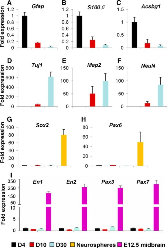
Figure 6.

Expression analysis of molecular markers of iN cells. A–H, mCherry+ cells in the dorsal midbrain derived from WT mice that were infected with the virus AAV–Ascl1/mCherry at P12–P15 were collected by FACS on day 4 (D4, black bars), day 10 (D10, red bars), and day 30 (D30, blue bars) after infection. The expression of the astrocyte markers Gfap (A), S100β (B), and Acsbg1 (C), the neuronal markers Tuj1 (D), Map2 (E), and NeuN (F), the neural progenitor markers Sox2 (G) and Pax6 (H), and the midbrain neural progenitor markers En1, En2, Pax3, and Pax7 (I) was examined by qRT-PCR. The neurospheres (yellow bars) derived from the SVZ of mice at P0 were used as positive controls for detecting the expression of Sox2 and Pax6. The cells derived from E12.5 midbrain (purple bars) were used as positive controls for detecting the expression of En1, En2, Pax3, and Pax7.
