Figure 2.
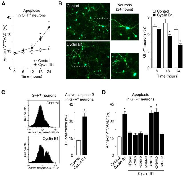
Cyclin B1 induces neuronal apoptotic death via the intrinsic mitochondrial pathway activation. A, Rat cortical neurons were transfected with 0.8 μg/106 cells pIRES2–EGFP, either empty (Control; expressing GFP) or containing the full-length cDNA of human cyclin B1 (Cyclin B1; expressing both cyclin B1 and GFP) for 6–24 h. cyclin B1 expression time dependently induced neuronal apoptotic death, as assessed by annexin V+/7-AAD− quantification by flow cytometry. *p < 0.05 versus control. B, Epifluorescence microphotographs revealed the typical axonal disruption of apoptosis in cyclin B1-transfected neurons (24 h). Scale bar, 50 μm. Transfection rate (percentage of GFP+ neurons) was determined by flow cytometry. *p < 0.05 versus control. C, Cyclin B1 induced caspase 3 cleavage and activation by 24 h after transfection of neurons, as showed by quantitative analyses of GFP+ (transfected) neurons by flow cytometry. *p < 0.05 versus control. D, Rosc (10 μm) or caspase inhibitors (zVAD-fmk and zDEVD-fmk; 100 μm) prevented cyclin B1-mediated neuronal death at 24 h of transfection. Inhibition of caspases-8 (zIETD-fmk; 100 μm) or caspase-2 (zVDVAD-fmk, 100 μm) was ineffective. Caspase-3 (zDQMD-fmk; 100 μm) or caspase-9 inhibition (zLEHD-fmk; 50 μm) abrogated cyclin B1-mediated neuronal death. *p < 0.05 versus control. In all cases, data are the mean ± SEM of four independent neuronal cultures (n = 4).

