
Figure 1.
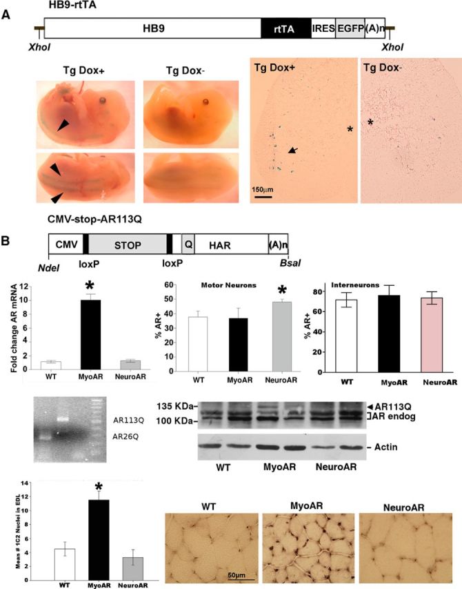
A, Top, Diagram of HB9-rtTA-IRES-EGFP transgene. HB9, Ppromoter of homeobox9 gene; rtTA, reverse tet-controlled transactivator; IRES, internal ribosomal entry site; EGFP, enhanced green fluorescent protein; (A)n, mouse metallothionein polyadenylation signal. Bottom, Photomicrographs of crosses of LacZ reporter mice with HB9-rtTA-IRES-EGFP/tetO-Cre mice. Bottom left, E14.5 Tg embryos gestating in dams treated with the tetracycline analog Dox for 3 d before dissection (Dox+) or untreated dams (Dox−). Arrows indicate staining along spinal column corresponding to ventral roots. Bottom right, Sections from adult Tg mice either treated with Dox or untreated demonstrating staining in motor neurons after Dox administration. Asterisk indicates the position of central canal. Arrow indicates stained motor neurons after Dox treatment. Scale bar, 150 μm. B, Top, Diagram of CMV-AR113Q transgene. CMV, mini cytomegalovirus promoter; STOP, floxed NEO-stop cassette; HAR, human AR clone with expanded CAG repeat (Q). loxP sites and restriction sites used to excise the constructs from plasmid before microinjection are also indicated. Second row, AR measurement in MyoAR and NeuroAR mice. As expected, AR was elevated in muscle only in MyoAR mice and in motor neurons only in NeuroAR mice. Left, qPCR measurement of AR in anterior tibialis muscle indicates AR mRNA is elevated in myoAR mice ∼10× relative to WT (n, 6/group). Middle, the percentage of motor neurons immunoreactive for AR in the retrodorsolateral nucleus is elevated relative to WT (n = 6–11 per group). Right, No differences in the number of AR+ interneurons in lamina X (n = 3–5 per group). *p < 0.05 for indicated comparison. Third row, PolyQ-expanded AR is expressed selectively in muscle. Left, PCR products obtained using primers that flank the CAG repeat region of the transgene on genomic DNA from CMV-stop-AR113Q mice demonstrate expanded region relative to DNA from an AR26Q clone. No template control and 100 bp ladder are also shown. Right, Western blot for AR in muscle of two mice of each genotype showing that polyQ AR (AR113Q) is detectable only in MyoAR mice. Actin Western blot used as a loading control is shown at bottom. Fourth row, 1C2-immunoreactive nuclei are observed in muscle of MyoAR mice. Left, Graph indicating that the number of 1C2+ nuclei is increased selectively in MyoAR mice (n = 3/group). *p < 0.05 for indicated comparison. Right, Photomicrographs illustrating increased 1C2 staining in EDL muscle of MyoAR mice. Scale bar, 50 μm.