
Figure 4.
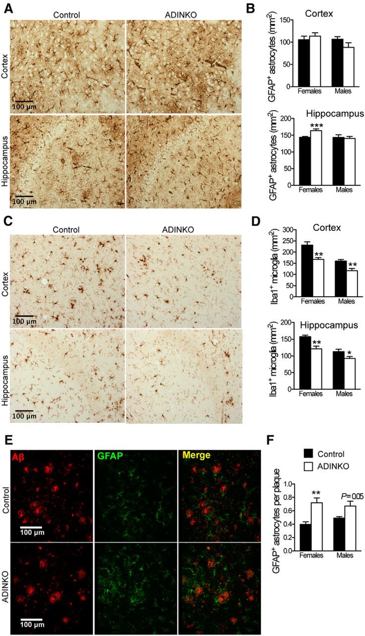
ADINKO brains show less microgliosis with astrocytes accumulating in amyloid plaque vicinity. A, Representative anti-GFAP IHC for astrocyte analysis. B, GFAP-positive astrocytes were more abundant in ADINKO female hippocampus, but not in motor cortex. C, Representative IHC of Iba1-positive microglia. D, Infiltration of Iba1-positive microglia was reduced in ADINKO motor cortex and hippocampus. Males had fewer microglia than females. E, Representative micrograph with plaques (red) and GFAP-positive astrocytes (green) accumulating close to plaques. F, More GFAP-positive astrocytes accumulated around plaques in ADINKO cortex. Student's t test, n = 7 per group, *p < 0.05, **p < 0.01, mean ± SEM.