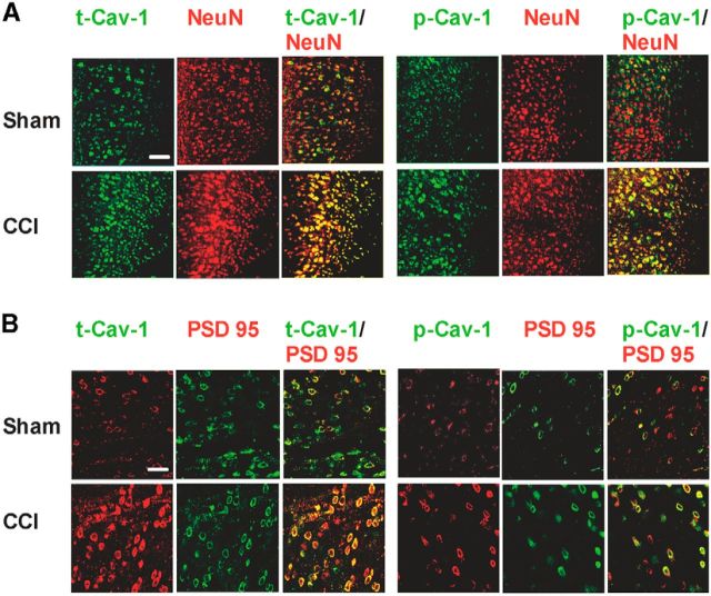
Figure 2.
Cellular and subcellular localization of t-Cav-1 and p-Cav-1 expression in the ACC. A, Double immunofluorescence showed that t-Cav-1 and p-Cav-1 expression in contralateral ACC were increased and expressed in neurons in CCI mice (colocalization with NeuN; n = 4). Scale bar, 100 μm. B, The increased t-Cav-1 and p-Cav-1 colocalized with PSD-95, a postsynaptic marker (n = 5). Scale bar, 50 μm.

