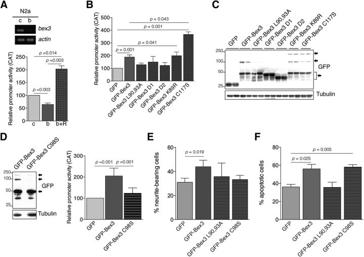
Figure 6.
Bex3 dimerization potentiates the transcriptional activity of the mouse trkA promoter. A, Bex3 depletion reduces mouse trkA promoter activity. Top, Experiments using N2a cells were performed as described in Figure 3A upon infection with lentivirus-expressing control shRNA (c) and mouse Bex3 shRNA (b). Bottom, N2a cells were transfected with the reporter construct pMS8 together with control shRNA (c), Bex3 shRNA (b), or Bex3 shRNA + GFP-Bex3-resistant (R) expression vectors. Data are shown as the mean ± SEM (100% vs 64.3 ± 5.6%, control shRNA vs Bex3 shRNA, respectively); 100% vs 203 ± 12.5%, control shRNA vs Bex3 shRNA + GFP-Bex3-resistant expression vectors, respectively; n = 3–5 using duplicates). The p values were calculated using a paired two-tailed Student's t test. B, GFP-Bex3 expression increases the transcriptional activity of the mouse trkA promoter in N2a cells. Data are shown as the mean ± SEM (100% vs 188.7 ± 16.9%, GFP vs GFP-Bex3, respectively; 188.7 ± 16.9% vs 366 ± 20.5%, GFP-Bex3 vs GFP-Bex3 C117S, respectively; n = 4–9 using duplicates). The p values were calculated using a paired two-tailed Student's t test. Note the increased trkA promoter activity induced by GFP-Bex3, GFP-Bex3 K89R, and GFP-Bex3 C117S protein, and the lack of effect on the trkA promoter upon GFP-Bex3 mutants L90,93A, D1, and D2 expression. C, Representative Western blot showing GFP-Bex3 protein expression in N2a transfected cells, in duplicate. Note the presence of monomers, dimers, and tetramers of GFP-Bex3 proteins, indicated by arrows. D, Bex3 dimerization is required to enhance the transcriptional activity of the trkA promoter. Western blot showing the expression of WT Bex3 and Bex3 C98S, which does not dimerize in N2a cells (left). Reporter assays were performed as described in A (right). Data are shown as the mean ± SEM (205 ± 15.25% vs 123.6 ± 10.34%, GFP-Bex3 vs GFP-Bex3 C98S, respectively; n = 5 using duplicates). The p values were calculated using a paired two-tailed Student's t test. Arrows indicate GFP-Bex3 monomers, dimers, and tetramers. E, Bex3 overexpression increases neurite outgrowth of PC12 cells in response to NGF. Cells were transfected with plasmids expressing GFP, GFP-Bex3, GFP-Bex3 L90,93A, and GFP-Bex3 C98S, and the next day were stimulated with NGF (10 ng/ml) for 96 h. Neurite outgrowth was quantified as described in Figure 3D. Data are shown as the mean ± SEM. The percentage of neurite-bearing cells versus the total number of cells scored in each case was 31.1% and 376 cells for GFP, 43.9% and 275 cells for GFP-Bex3, 35.7% and 281 cells for GFP-Bex3 L90,93A and 33.2% and 230 cells for GFP-Bex3 C98S. The p value was calculated using a paired two-tailed Student's t test. F, Overexpression of Bex3 protein induces apoptosis of NGF-dependent sensory neurons in vitro. Cultured DRG neurons were transfected with plasmids expressing GFP, GFP-Bex3, GFP-Bex3 L90,93A, and GFP-Bex3 C98S. Apoptotic neurons were quantified as described in Figure 3C. Data are shown as the mean ± SEM. The percentage of apoptotic cells versus the total number of cells scored in each case was 36.0% and 257 cells for GFP, 56.0% and 127 cells for GFP-Bex3, 35.6% and 142 cells for GFP-Bex3 L90,93A, and 58.1% and 120 cells for GFP-Bex3 C98S. The p value was calculated using a paired two-tailed Student's t test.

