Figure 5.

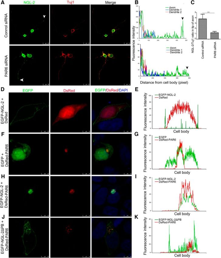
NGL-2 is recruited by PAR6 and polarized distribution at the tip of axon. A, The effect of the knockdown of PAR6 on NGL-2 distribution. Hippocampal neurons were transfected with PAR6 siRNA or control siRNA. Arrowhead indicates the tip of the axon. Scale bar, 25 μm. B, Profiles of NGL-2 immunofluorescence intensity in the processes of stage 2 and stage 3 neurons. Fluorescence intensities were plotted against the distance from soma, as shown in A. C, NGL-2/Tuj1 ratios in axon tips of stage 3 neurons. The NGL-2/Tuj1 ratio in the axon tip of neurons transfected with control siRNA was 3.81 ± 0.89 (n = 50). The NGL-2/Tuj1 ratio in the axon tip of neurons transfected with PAR6 siRNA was 1.2 ± 0.26 (n = 50). The error bars represent the SD. ***p < 0.001, Student's t test. D–I, PAR6 recruits NGL-2 to one side of the HEK 293 cells. HEK 293 cells were transfected with EGFP-NGL-2 without or with DsRed-PAR6. After 48 h, cells were fixed. Scale bar, 10 μm. The immunofluorescence intensity of EGFP-NGL-2 and DsRed-PAR6 signals were measured by ImageJ. J, K, HEK 293 cells were cotransfected with EGFP-NGL-2▵PB and DsRed-PAR6. After 48 h, cells were fixed. Scale bar, 10 μm. The immunofluorescence intensity of NGL-2 and PAR6 signals was measured by ImageJ.
